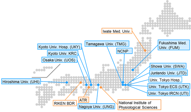
・Institutes in the blue boxes: Measurement and analysis sites for neuropsychiatric disorders
・Institute in the orange boxes: Show analysis support sites
・Institutes listed in boxes with a colored background: Participation in the traveling subject measurement
| Site | Role |
MRI
(System version)
|
Protocol | Main target population |
|---|---|---|---|---|
| The University of Tokyo ECS (UTK) | Data acquisition, Analysis | Prisma (VE11C) | CRHD | Adolescent cohort, HP, ASD, Sch, MDD, Epilepsy |
| The University of Tokyo IRCN (UTI) | Data acquisition, Sharing | Prisma (VE11C) | CRHD | HP, ASD, Sch, MDD, BPD |
| Advanced Telecommunications Research Institute International (ATR) | Data acquisition, Sharing, Analysis | Prisma (VE11C) | CRHD | HP |
| Fukushima Medical University (FUM) | Data acquisition | Skyra (VE11C) | HARP | HP, AD, PD |
| Tamagawa Academy & University (TMG) | HARP setup, Data acquisition | Trio (VB19A) | HARP | Adolescent cohort |
| Showa University (SWA) | HARP setup ,Data acquisition | Skyra (VE11E) | HARP | HP, ASD |
| National Center of Neurology and Psychiatry (NCNP) | HARP setup ,Data acquisition, Sharing, Analysis | Skyra (VE11E) | HARP | HP, Sch, MDD, AD, PD |
| Juntendo Hospital (JTD) | Data acquisition | Prisma (VE11C) | HARP | HP, PD, MSA, PSP |
| Osaka University (UOS) | Data acquisition | Prisma (VE11C) | HARP | HP, Chronic pain |
| Hiroshima University (UHI) | HARP setup, Data acquisition | Skyra (VE11C) | HARP | HP, MDD, BPD |
| Nagoya University (UNG) | Data acquisition | Verio (VB17A) | HARP | HP, Sch |
| Kyoto University (UKY) | HARP setup, Data acquisition | Skyra (VE11C) | HARP | HP, AD, PD |
| Kyoto University Kokoro Research Center (KRC) | Data acquisition | Verio (VB17A) | HARP | HP, Sch, MDD, BPD |
| RIKEN Center for Biosystems Dynamics Research (BDR) | HARP setup, Data Analysis | Prisma (VE11C) | HARP | NA |
※The data for UNG (Nagoya University) was obtained by the Brain Mapping by Integrated Neurotechnologies for Disease Studies (Brain/MINDS) project.
